Anatomy Textbook vs. Atlas
I’d like to share some of the advice I wish I’d known when I was in my…
I’d like to share some of the advice I wish I’d known when I was in my first year of medical school, things that, looking back, would have helped me tremendously.
For this post, I’ll focus just on the array of anatomy textbooks available to us.
1. My first piece of advice: don’t buy anything ahead of time.
Try out a few textbooks, see which one you like. They’re all different, they each have their advantages and disadvantages, and since everyone is different, everyone will tell you something different. So don’t invest in anything until you’ve had a chance to use them yourself. For advice on where to get cheap textbooks either hardcover or digital, send me a message and I’ll get back to you 🙂
2. Understand the difference between a textbook and an atlas:
Simply put, an atlas is a compilation of all the drawings of each region of the human body, with labels on each drawing. A textbook is a wordy, lengthy description of the body, which of course also has drawings included, but they’re usually not nearly as numerous or detailed as an atlas. There are some hybrid publications, such as Thieme, that I’ll discuss later. Generally, students pick one of each, although depending on your school, you might just end up using atlases or just textbooks…or just the Internet.
3. Remember: your #1 resource should be ~~~~LECTURES~~~~
If you can’t bring yourself to attend lecture, at least look at the lecture slides. Skim through them because they will help you focus the material. Anatomy is MASSIVE. No one will ever be able to learn it all. Use the lecture slides to guide you to the scope of knowledge you have to gain.
So, to begin: ATLASES: what are your options?
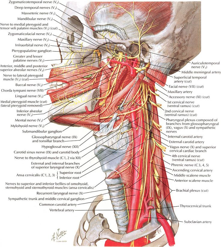
–Netter’s Atlas of Anatomy: This textbook came highly recommended by American med students. It has great drawings and almost always labels everything you could ever want to know about a picture (Gray’s, for example, does not do this, much to my annoyance). The muscle tables in the back were excellent for learning upper/lower limb insertions (I only discovered them after I spent ages making my own tables). Great for bones, joints, and internal organs, but the sheer number of labels can be overwhelming at times. I used Netter’s as a way to get the larger picture, but there were certain things that I had to look for elsewhere (ex. arteries and veins, and where they branch from).
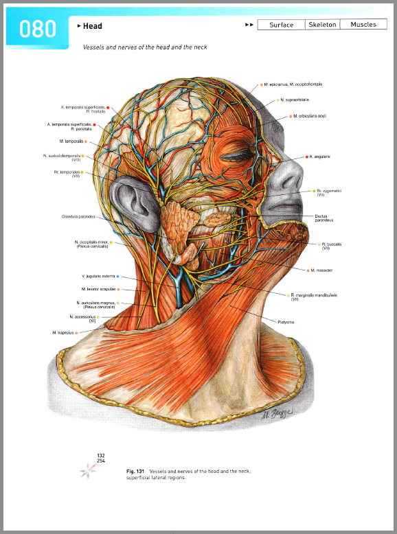
-Sobotta Atlas of Human Anatomy: similar to Netter’s, came highly recommended by European med students (beloved by my own teachers), and also has a lot of labels, but perhaps not to the extent that Netter’s does. Also, it annotates whether its an artery (red) or nerve (yellow), which helps for when you’re quizzing yourself. Sobotta I often found myself using for internal organs, mostly, and it had a lot of good cross-section images. (Also arteries, if I remember correctly, but I can’t find a good chart right now.)
BE VERY CAREFUL: there are two editions of Sobotta, one with English and one with Latin nomenclature, don’t buy the wrong one by accident cause you’ll be struggling (as I was when the library was all out of English editions).
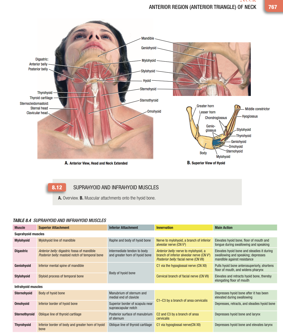
-Grant’s Atlas of Anatomy: I would say this is a sort of hybrid atlas-textbook, because it has charts showing muscle origins/insertions, nerve branchings and a horde of useful little factoids. The diagrams vary from being clear and to the point, like below, or all-encompassing, similar to Netter’s. The more I used Grant’s, the more I liked it. Sadly, I didn’t discover it in time, although our teachers often took diagrams from there for labeling quizzes. 😉
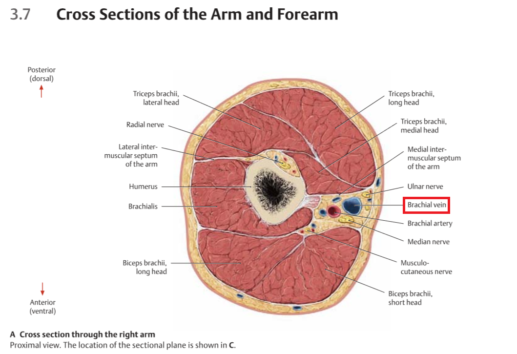
Thieme Atlas of Anatomy: called an atlas, but like I said, I’d classify it as a hybrid. It comes in a few volumes (musculoskeletal system, head/neck/neuroanatomy system, internal organs) and has so-called “big Thieme” and “little Thieme.” A lot of people swore by little Thieme (a pocketbook with tons of great information), but I couldn’t stand it. My main issue was that the images weren’t labeled with words, only numbers, and you had to find which number correlated to which structure on the other page. Check it out, see if you like it. Big Thieme refers to the full-sized textbooks that I loved. Thieme was a ton of people’s #1 resource for all regions of the body, including neuroanatomy. (Note: a lot of Thieme textbooks came with an online component, don’t be fooled. Neither me nor my friends ever got it to work :[ ). Wish I could have found a better picture to showcase it’s “hybrid-ness” but unfortunately this is the only kind of images I can find right now.
TEXTBOOKS:
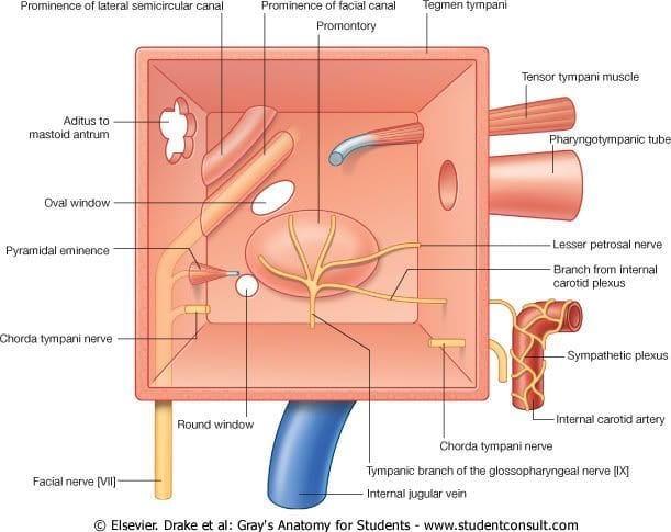
-Gray’s Anatomy for Students: excellent spatial representations of difficult-to-understand topics, like spaces/crevices of the body. This book is a combination of representative diagrams and very clear, focused anatomical drawings. Exceptional for visualizing the body. Could not use for neuroanatomy: simply not detailed enough for our school. I found that it was difficult/annoying to find concepts when I was trying to review a topic (i.e. the bladder was discussed across three different sections), but it was great for skimming/reading a chapter start to finish. Note: several of our teachers disliked Gray’s for Students, but I always felt like it was a good source to jump to when something wasn’t clear. It did a great job of isolating concepts to better understanding.
-Moore’s Clinically Oriented Anatomy: I absolutely loved it. I used it mostly for internal organs because it was succinct and went through each organ in each system in a clear, systematic format: structure, innervation, blood supply. Easy as a reference, easy to follow. Also useful for, believe it or not, upper and lower limbs. I found that the muscle insertions were a lot easier to memorize when you understood why and how and Moore’s, as its title suggests, gives relevant, interesting clinical information that makes the rather dry musculoskeletal system tons more interesting (at least for me). Moore’s, however, was also not detailed enough for neuroanatomy.
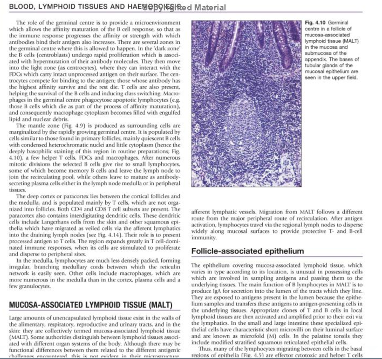
-Gray’s Anatomy: The Anatomical Basis of Clinical Practice 40th Edition (aka the “Bible”). Massive book, would not recommend reading front to back but this book was an absolute gem. Again, I discovered it much too late in my journey through med school to make proper use of it, but when I truly didn’t understand something and was at a loss for what to do, I knew I could open Gray’s and it would clear it right up. For example, understanding the inner ear was never clearer than when I read the few pages related to it in Gray’s. Gray’s (for Adults, as we’d affectionately called it) was amazing for histology, much to my surprise, because it gave a foundation, an understanding, to certain histological structures that, up until then, I had just been rote memorizing. (This blurry photo does not do this incredible textbook justice!)
BONUS: Color Atlas of Anatomy (Rohen/Yokochi): a one-of-a-kind book that is excellent for reviewing identification because it shows real photos of cadavers, beautifully dissected. I won’t post an image but definitely look it up, it’s an indispensable resourcefor a student preparing for a pin-type/gross anatomy oral test.
If you’re interested in other resources, check out my Resources Masterpost!
Hope this helps 🙂 If yes, I can offer advice on our other classes, including histology, physiology, biochemistry, etc. etc. And if you have any questions about the resources, where to get them at reasonable costs, or my medical school, shoot me a message. I’d love to hear from you. Fellow upper years, let me know if you think of anything I missed or have other advice to offer!
Copyright note: I do not own any of these pictures; they belong to the individual artist/publisher, and I did not intend any sort of copyright infringement in posting them here for reference.

